Xiaojuan Li, PhD; Thomas Link, MD, PhD
Department of Radiology and Biomedical Imaging
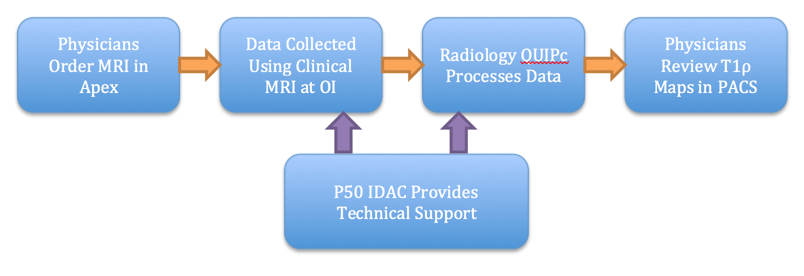
From Feb 2011 – Oct 2015, MR T1ρ imaging was added to 275 clinical cases at the Orthopaedic Institute (OI), UCSF, using the clinical 3T MRI, including 205 knee cases and 70 hip cases. The physicians can put in the MR order through the clinical ordering system. The data are collected during the clinical MRI scan, followed by data processing (including cartilage segmentation and the generation of color-coded T1ρ maps) by Radiology Quantitative Imaging Processing Center (QUIPc). The T1ρ maps are pushed back to the clinical PACS system which can be reviewed directly by physicians.
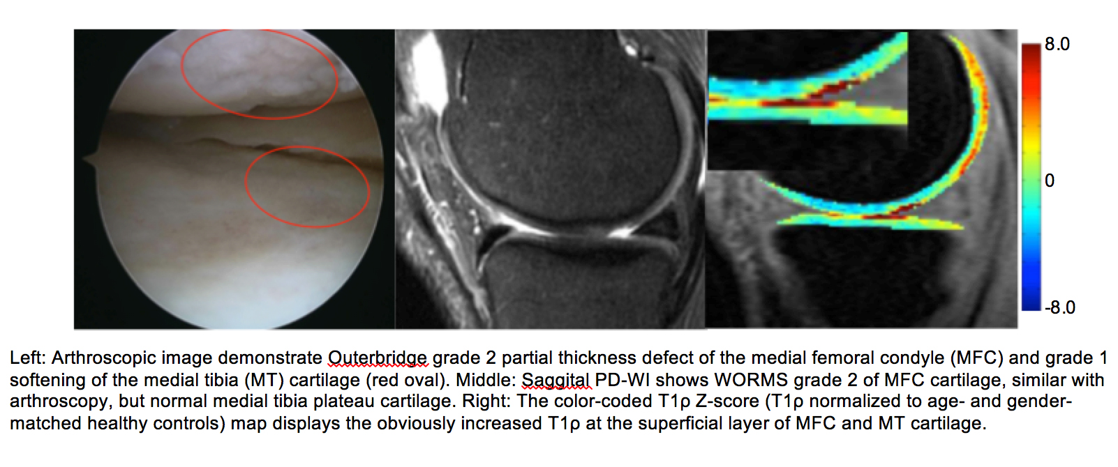
 We now use T1ρ quantitative MR imaging routinely for clinical use. Its ability for us to detect cartilage injuries before loss of tissue really enhances our ability to take care of athletes and their demand of their joints. It is a great diagnostic tool that has improved our patient care. T1ρ imaging allowed us to detect this cartilage injury that is not seen on conventional MRI (Figures below). We are able to provide the appropriate treatment to the patient and she has since recovered well. -C. Benjamin Ma, Chief, Sports Medicine and Shoulder Surgery, UCSF