Michael Samaan, PhD
UCSF Post-Doctoral Research Scholar
Department of Radiology and Biomedical Imaging
Data suggests reduced sensory perception at the MFC and RH, as well as trends toward altered lower extremity kinematics/kinetics and activity levels at baseline, in those that exhibit hip OA progression. VPT provides a quantitative assessment of sensory perception and may help researchers understand the relationship between sensory perception and the pathogenesis of hip OA.

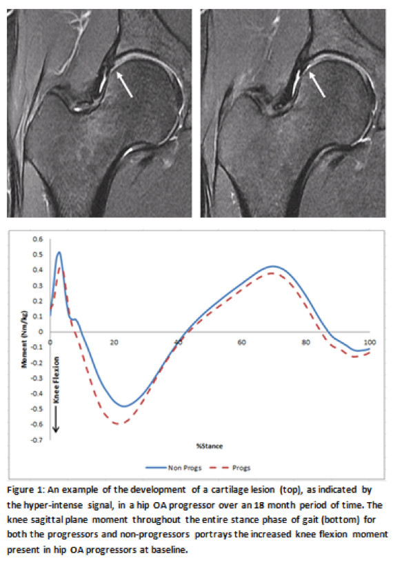
A total of 10 progressors were present in this cohort. Progressors possessed lower BMI compared to the non-progressors (p=0.01). Progressors trended towards reduced peak hip extension (p=0.09), increased peak knee flexion moment (p=0.06) and increased knee flexion moment impulse (p=0.09) compared to non-progressors. In addition, progressors demonstrated significantly decreased sensory perception at the MFC (p=0.02) and RH (p=0.02) as well as a trend towards decreased sensory perception at the MM (p=0.06). No differences in pain scores, moderate or walking activity levels exist between the groups yet vigorous activity levels were trending towards significance (p=0.06) with progressors performing more vigorous types of activities such as running.

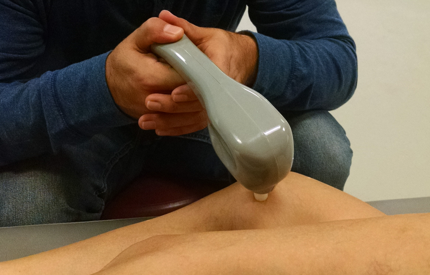
Figure 2: Vibratory perception testing (VPT) is performed using a biothesiometer, consisting of a
vibrating head that is placed at various anatomical landmarks. VPT allows researchers to assess
sensory perception as an effect of various pathologies including hip osteoarthritis.